படிமம்:Hepatitis B virus v2.svg

மூலக்கோப்பு (SVG கோப்பு, பெயரளவில் 512 × 350 பிக்சல்கள், கோப்பு அளவு: 11 KB)
| இது விக்கிமீடியா பொதுக்கோப்பகத்தில் இருக்கும் ஒரு கோப்பாகும். இக்கோப்பைக் குறித்து அங்கே காணப்படும் படிம விளக்கப் பக்கத்தை இங்கே கீழே காணலாம்.
|
உள்ளடக்கம்
சுருக்கம்
| விளக்கம்Hepatitis B virus v2.svg |
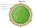
English: Simplified graphical representation of a cross-section of the Hepatitis B virus particle and surface (surplus) antigen, the hepatitis B e antigens (HBcAg) shown are considered not part of the viral particle (quod vide viral nonstructural protein). The structure of the Hepatitis B virus as first described by Dane & al.[1] and Jokelainen, Krohn & al.[2] during 1970. The hepatitis B virion is a complex, double shelled, spherical particle with a 42 nm diameter.[1][2][3]
The virion was initially referred to as the Dane particle.[4] Only after Baruch Blumberg received the Nobel Prize in Medicine during 1976 was it universally accepted that the particle is a virus and the infectious agent of Hepatitis B.
|
| நாள் | |
| மூலம் | Own work based on: Hepatitis B virus v2.png 14 நவம்பர் 2007 (original upload date) Created by en:User:GrahamColm. Original uploader was TimVickers at en.wikipedia |
| ஆசிரியர் | vectorization Glrx |
| ஒத்தக்கோப்பு |
[தொகு] |
| SVG genesis InfoField | This vector image was created with a text editor. |
அனுமதி
| This file is made available under the Creative Commons CC0 1.0 Universal Public Domain Dedication. | |
| The person who associated a work with this deed has dedicated the work to the public domain by waiving all of their rights to the work worldwide under copyright law, including all related and neighboring rights, to the extent allowed by law. You can copy, modify, distribute and perform the work, even for commercial purposes, all without asking permission.
http://creativecommons.org/publicdomain/zero/1.0/deed.enCC0Creative Commons Zero, Public Domain Dedicationfalsefalse |
எல்லா மொழிபெயர்ப்புகளும்
Translations added to this section should be free of copyright claims (either CC0 or public domain).
- Hepatitis B core antigen (HBcAg) ≅ Hepatitis core antigen (Q24723361)
- Hepatitis core antigen (Q24723361)
- Hepatitis B e antigen ≅ Hepatitis B e antigen (Q75838622)
- Hepatitis B e antigen (Q75838622)
- Hepatitis B surface antigen (HBsAg) ≅ HBsAg (Q2282887)
- HBsAg (Q2282887)
- DNA polymerase ≅ DNA polymerase (Q206286)
- டி. என். ஏ பாலிமரேசு (Q206286)
- Partially double-stranded DNA ≅ DNA (Q7430)
- டி. என். ஏ. (Q7430)
Sources
- ↑ a b c D.S. Dane , C.H. Cameron , Moya Briggs (1970). "Virus-Like Particles in Serum of Patients with Australia-Antigen-Associated Hepatitis". The Lancet 295: 695–698. DOI:10.1016/S0140-6736(70)90926-8.
- ↑ a b c d e f g h i j k l P. T. Jokelainen, Kai Krohn, A. M. Prince and N. D. C. Finlayson (1970). "Electron Microscopic Observations on Virus-Like Particles Associated with SH Antigen". Journal of Virology 6 (5): 685-689. ISSN 1098-5514.
- ↑ a b c d e f The hepatitis B virus. World Health Organisation.
- ↑ a b Almeida J D, Rubenstein D & Scott E J. (1971). "New antigen-antibody system in Australia-antigen-positive hepatitis". The Lancet 298 (7736): 1225–7. DOI:10.1016/S0140-6736(71)90543-5.
- ↑ Bayer, M. E., B. S. Blumberg, and B. Werner (1968). "Particles associated with Australia antigen in the sera of patients with leukemia, Down's syndrome and hepatitis.". Nature (London) 218: 1057-1059.
- ↑ Baruch S. Blumberg, Harvey J. Alter, and Sam Visnich (Jul 1984). "Landmark article Feb 15, 1965: A 'new' antigen in leukemia sera. By Baruch S. Blumberg, Harvey J. Alter, and Sam Visnich". Journal of the American Medical Association 252 (2): 252–7. DOI:10.1001/jama.252.2.252. PMID 6374187. ISSN 0098-7484.
- ↑ Prince, A. M. (1968). "An antigen detected in the blood during the incubation period of serum hepatitis". Proceedings of the National Academy of Science U.S.A. 60: 814-821.
Captions
Items portrayed in this file
சித்தரிப்பில் உள்ளது
Creative Commons CC0 License ஆங்கிலம்
30 சூலை 2024
கோப்பின் வரலாறு
குறித்த நேரத்தில் இருந்த படிமத்தைப் பார்க்க அந்நேரத்தின் மீது சொடுக்கவும்.
| நாள்/நேரம் | நகம் அளவு சிறுபடம் | அளவுகள் | பயனர் | கருத்து | |
|---|---|---|---|---|---|
| தற்போதைய | 12:45, 1 ஆகத்து 2024 |  | 512 × 350 (11 KB) | Glrx | Uploaded own work with UploadWizard |
கோப்பு பயன்பாடு
இப் படிமத்துக்கு இணைக்கப்பட்டுள்ள பக்கங்கள் எதுவும் இல்லை.
மேனிலைத் தரவு
இந்தக் கோப்பு கூடுதலான தகவல்களைக் கொண்டுளது, இவை பெரும்பாலும் இக்கோப்பை உருவாக்கப் பயன்படுத்திய எண்ணிம ஒளிப்படக்கருவி அல்லது ஒளிவருடியால் சேர்க்கப்பட்டிருக்கலாம். இக்கோப்பு ஏதாவது வகையில் மாற்றியமைக்கப்பட்டிருந்தால் இத்தகவல்கள் அவற்றைச் சரிவர தராமல் இருக்கலாம்.
| குறுகிய தலைப்பு | Hepatitis B virus v2 |
|---|---|
| அகலம் | 100% |
| உயரம் | 100% |


